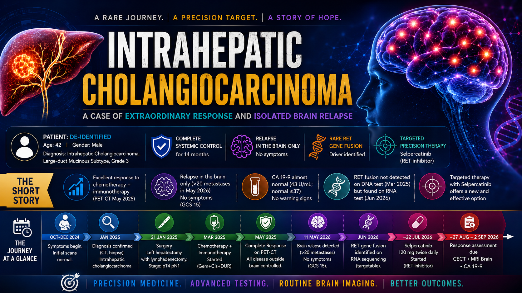
- Core Finding: Durable systemic control on chemo-immunotherapy does not preclude silent, high-burden CNS-only relapse in iCCA, often with near-normal tumor markers like CA 19-9.
- Diagnostic Impact: DNA+RNA profiling of metastatic tissue is critical for detecting actionable fusions (e.g., NCOA4::RET), especially when prior DNA-only assays are negative or limited by cellularity.
- Actionable Protocol: Following surgery and WBRT, CNS-penetrant RET inhibitors (e.g., selpercatinib) offer a tumour-agnostic, targeted therapy option for RET fusion-positive iCCA with CNS metastases.
TL;DR
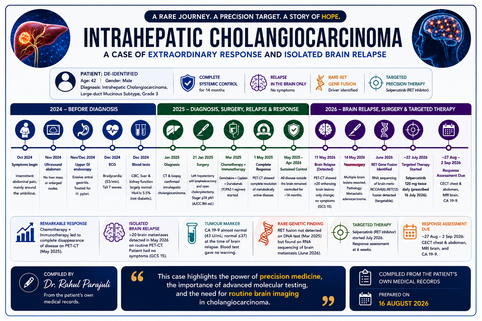
- CNS-only relapse occurred after ~14 months of excellent extracranial control on gemcitabine–cisplatin + durvalumab.
- >20 brain metastases were found on surveillance imaging without neurologic symptoms and with near‑normal CA 19‑9.
- Actionable biology (NCOA4::RET fusion) emerged only after DNA+RNA profiling of a resected brain metastasis (DNA-only assays were negative/insufficient).
- Management pivoted to CNS‑penetrant RET inhibition (selpercatinib) after surgery + WBRT.
Title
Intrahepatic cholangiocarcinoma with CNS-only relapse and an actionable NCOA4::RET fusion: a de-identified case report
Abstract
High‑risk resected intrahepatic cholangiocarcinoma relapsed early post‑op but then achieved deep systemic control on gemcitabine–cisplatin plus durvalumab. Despite ~14 months of extracranial control, surveillance imaging uncovered multifocal intracranial metastases (>20) with no neurologic symptoms and near‑normal CA 19‑9. A resected brain metastasis underwent DNA+RNA profiling, revealing an NCOA4::RET fusion not seen on prior DNA‑only testing, prompting selpercatinib. Key takeaways: CNS‑only relapse can be silent; tumour markers can underperform for intracranial progression; and RNA fusion testing can be management‑changing when DNA results are negative or limited.
Background
CNS involvement in cholangiocarcinoma is uncommon and may be clinically silent; evidence for CNS‑directed strategy is largely case‑based.[1][2] RET fusions are rare but actionable with tumour‑agnostic RET inhibitors; RNA-inclusive profiling can be critical for fusion detection.[3]
Case presentation
- Patient: adult (early 40s), ~75 kg
- Presenting symptom (initial): intermittent abdominal pain
- Risk factors: no documented viral hepatitis; fatty liver and hypertriglyceridemia noted in prior testing
- Initial tumour marker: CA 19-9 markedly elevated at diagnosis
Investigations
Imaging (key studies)
- Initial cross-sectional imaging: intrahepatic lesion (~4 cm) with satellite lesions and suspicious abdominal lymphadenopathy
- Baseline PET-CT: FDG-avid hepatic disease and nodes; CNS reported without abnormal uptake (MRI preferred for CNS)
- Postoperative PET-CT (~7 weeks): peritoneal/omental disease with ascites consistent with progression
- Response assessment: PET-CT later showed complete metabolic response
- Relapse detection: surveillance PET-CT demonstrated new multifocal intracranial lesions with otherwise stable extracranial findings
- MRI brain: multiple enhancing supra- and infratentorial lesions with associated edema; some hemorrhagic features reported
Pathology
- Surgery specimen: large-duct mucinous intrahepatic cholangiocarcinoma, grade 3; multifocal; LVI/PNI present
- Nodal status: 5/5 nodes positive; pathologic stage pT4 pN1 (AJCC 8)
- Brain lesion: metastatic adenocarcinoma consistent with the known primary (IHC supportive)
Laboratory and biomarkers
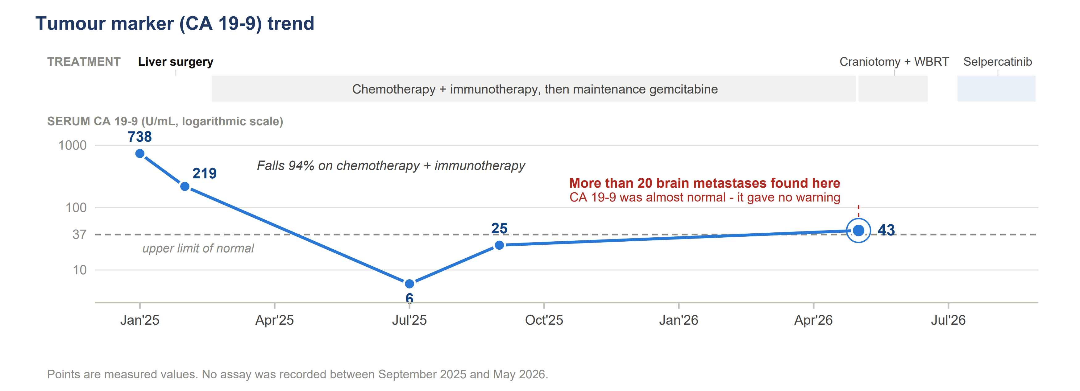
- CA 19-9 trend (selected): very high at diagnosis → substantially decreased on therapy; near-normal at time of CNS relapse
- Safety-relevant baseline issues from available records: bradycardia/borderline QTc; thrombocytopenia reported at one point; mild transaminitis; hepatic steatosis and prior hepatectomy
Therapeutic interventions
Local therapy
- Hepatic surgery: left hepatectomy with additional hepatic resection, portal lymphadenectomy, and cholecystectomy
- Neurosurgery: resection of the largest intracranial lesion
- Radiation: whole-brain radiotherapy (10 fractions; total dose not documented in available records)
Systemic therapy
- First-line systemic therapy: gemcitabine + cisplatin + durvalumab (TOPAZ-1 regimen), followed by ongoing systemic therapy/maintenance per records.[4]
- Targeted therapy: selpercatinib initiated after RET fusion detection (dose per prescription in records)
Molecular profiling (why RNA mattered)
Three assays in the record yielded progressively more actionable information:
- Liquid biopsy (DNA-only): no clinically significant pathogenic variants reported
- Primary-tumour tissue profiling (DNA-based): alterations reported but RET not identified (noted low tumour cellularity)
- Brain metastasis profiling (DNA+RNA): NCOA4::RET fusion detected with high expression
Interpretation: DNA-only panels can miss gene fusions; RNA-based fusion calling can materially change management.
Outcome and follow-up (as documented)
As of mid‑Aug 2026, the patient had completed ~4 weeks of selpercatinib with planned reassessment at ~6 weeks (CECT chest/abdomen, MRI brain, CA 19‑9). Radiographic response after RET inhibition was not available in the provided records.
Discussion
This case raises several practice-relevant issues:
- CNS sanctuary relapse: durable extracranial control does not exclude clinically meaningful intracranial progression; symptom absence does not rule out high CNS tumour burden.
- Tumour markers may mislead: CA 19-9 was only minimally elevated at CNS relapse; biomarker trends should not replace imaging when clinical risk is high.
- Test selection matters: the actionable RET fusion emerged only after DNA+RNA profiling of metastatic tissue. Low tumour cellularity and the biological nature of fusions can limit DNA-only sensitivity.
- Targeted therapy rationale: selpercatinib is tumour-agnostic for RET fusion-positive solid tumours and is a reasonable choice when CNS penetration is clinically important.
- This rationale is supported by tumour-agnostic efficacy data for RET fusion-positive solid tumours (LIBRETTO-001).[3]
Learning points
- “Good systemic control” ≠ “no CNS risk”—CNS‑only progression can be clinically silent.
- CA 19‑9 may be near‑normal during intracranial progression—don’t over‑rely on biomarkers.
- If suspicion remains high, upgrade to DNA+RNA (especially with low cellularity or prior negative DNA).
- RET inhibitors offer tumour‑agnostic options where CNS activity matters.
Ethics, consent, and de-identification
- Consent: Written informed consent taken from the patient before compilation. This write-up is de-identified for only educational purposes.
- Identifiers removed: name, exact date of birth, precise residence, hospital IDs/registration numbers.
- Data availability: based on a compiled record set; primary source reports should be reviewed for clinical decisions.
References
- Chindaprasirt J, Sookprasert A, Sawanyawisuth K, et al. Brain metastases from cholangiocarcinoma: a first case series in Thailand. Asian Pacific Journal of Cancer Prevention. 2012. https://pubmed.ncbi.nlm.nih.gov/22901160/
- Oh D‑Y, He AR, Qin S, et al. Durvalumab plus gemcitabine and cisplatin in advanced biliary tract cancer (TOPAZ‑1). NEJM Evidence. 2022. https://evidence.nejm.org/doi/full/10.1056/EVIDoa2200015
- Xie P, Song Q, Shi H, Xiao Y. Case report: immunotherapy successfully treated brain metastasis in intrahepatic cholangiocarcinoma and literature review. Frontiers in Oncology. 2022. https://doi.org/10.3389/fonc.2022.911202
- Subbiah V, Wolf J, Konda B, et al. Tumour-agnostic efficacy and safety of selpercatinib in patients with RET fusion-positive solid tumours other than lung or thyroid tumours (LIBRETTO‑001). The Lancet Oncology. 2022. https://doi.org/10.1016/S1470-2045(22)00541-1
Academic Collaboration & Consulting
Select a clinical research or writing service below to inspect deliverables and submit an immediate, direct inquiry to Dr. Rahul Parajuli.
Article Review & Literature Review
Rigorous scientific evaluation of medical literature and manuscript drafts. Providing structured peer-review quality assessments, methodology audits, and exhaustive literature mapping for prospective studies or clinical guidelines.
Key Deliverables & Methodologies:
- Critical Appraisal & Peer Review Reports
- Systematic Literature Search & Evidence Mapping
- PRISMA-compliant Literature Reviews
- Methodological Quality & Risk of Bias Assessment

Dr. Rahul Parajuli
Physician & ResearcherPhysician, Clinical Informatics Researcher & Health-AI Architect. Specializes in acute critical care telemetry, full-stack biomedical engineering, Schema entity graph architectures, and high-performance search systems.

Community Discussion
0 Contributions